
eISSN: 1937-8688

Keloid: a rare clinical image

Mayank Rai, Sadhana Misar Wajpeyi

PAMJ. 2024; 49:97. Published 27 Nov 2024 | DOI: 10.11604/pamj.2024.49.97.45647
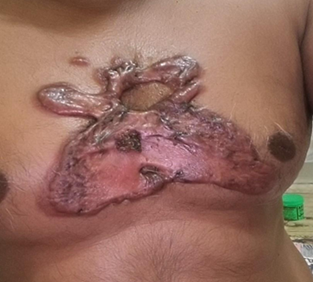
Keloid: a rare clinical image
At the location of a healed skin injury, keloids are an overgrowth of granulation tissue (collagen type 3), which is gradually replaced by collagen type 1. Keloids are glossy, fibrous nodules or firm, rubbery lesions that can range in color from red to black or pink to the color of the person´s skin Brown in hue. The average keloid scar is greater than the actual wound. They might take weeks or months to develop completely. They´re most commonly found on the chest, shoulders, earlobes, and cheeks. A 40-year-old male came with complaints of a lumpy or ridged area of skin with a pinkish color that was painless along with itching on the chest region for 8-9 months. On clinical examination and microscopic examination, a biopsy was done. The patient had normal blood count and glucose levels. Through this, it was diagnosed as keloid. As per Ayurved local application of alkaline preparation was given and the patient was kept on conservative treatment for 3 months. After 1 month of follow-up, there was no itching and the keloid became dry, and blackish with a size reduction. This image could be useful for differential diagnosis between hypertrophic scar, dermatofibromas, and keloid.

Corresponding Author

Mayank Rai, Department of Kayachikitsa, Mahatma Gandhi Ayurveda College, Hospital and Research Centre, Salod (H), Datta Meghe Institute of Medical Sciences (DU), Sawangi, Wardha, India (nrmankumayank@gmail.com)

Article Formats

Articles published in Pan African Medical Journal are Open Access and distributed under the terms of the Creative Commons Attribution 4.0 International (CC BY 4.0).

Open Access Logo

The Pan African Medical Journal (ISSN: 1937-8688) is a product of the PAMJ. The contents of this journal is intended exclusively for professionals in the medical, paramedical and public health and other health sectors.